The Vital Role of Plastic Surgeons in Multidisciplinary Care for Wound Healing After Endovascular Revascularization in Patients With Chronic Limb-Threatening Ischemia in Japan
Masahiko Fujihara, MD1; Mitsuyoshi Takahara, MD, PhD2; Osamu Iida, MD3; Yoshimitsu Soga, MD, PhD4; Terutoshi Yamaoka, MD, PhD5; Makoto Utsunomiya, MD, PhD6; Yoshiaki Yokoi, MD, PhD1
ISSN: 2694-3026
J CRIT LIMB ISCHEM 2022;2(1):E1-E7. Epub 2021 December 8.
Abstract
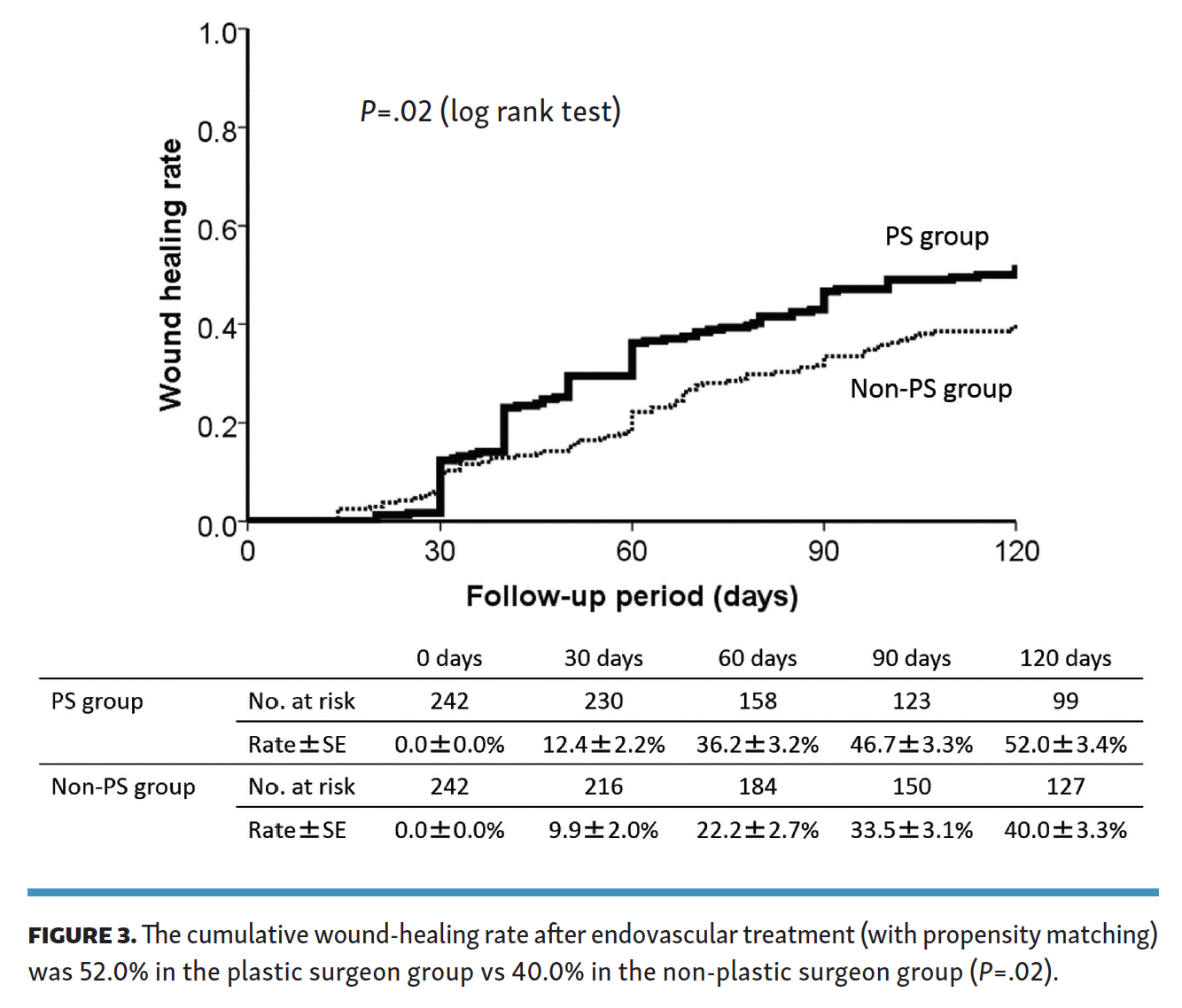
Objectives. Our study aim was to investigate the complete wound-healing rate in patients with chronic limb-threatening ischemia (CLTI) after endovascular revascularization, comparing the effectiveness of wound care management by plastic surgeons with other medical specialists. Methods. A retrospective review was conducted on 1000 CLTI cases of successful revascularization for de novo infrapopliteal lesions. At 8 hospitals, wound care management was performed by a plastic surgeon (PS group; n = 622 cases), and at 6 sites, wound care management was provided by other medical specialists (non-PS group; n = 244 cases). Propensity-score analysis was used for risk adjustment (n = 242 in each group). The primary outcome was the rate of complete wound healing at 4 months. Results. The PS group had a significantly higher wound-healing rate compared with the non-PS group (P=.02); the rate at 4 months was 52.0% vs 40.0%, respectively. Conclusion. Wound care management after successful revascularization by plastic surgeons showed a higher rate of wound healing compared with other specialists. Plastic surgeons in multidisciplinary team care provide the vital role of complete healing in CLTI treatment.
J CRIT LIMB ISCHEM 2022;2(1):E1-E7. Epub 2021 December 8.
Key words: chronic limb-threatening ischemia, endovascular therapy, peripheral arterial disease, plastic surgeon, wound care management
Introduction
Chronic limb-threatening ischemia (CLTI) is defined as extremity pain at rest or impending tissue loss caused by severely or chronically compromised blood flow. Above all, in patients with CLTI, the annual mortality rate and major amputation is high.1 Revascularization, including bypass surgery and endovascular treatment (EVT), is the standard theoretical option according to the latest guidelines.2-4 Several recent reports have also favored the efficacy of EVT for the initial treatment of CLTI patients and resulted in an annual decrease in major amputations, complete wound healing, and a reduced mortality rate.5-10 However, some patients did not benefit from successful revascularization concerning amputation-free survival and complete wound healing, and poor healing was reported in 20%-30% of cases.1,6
In recent years, several consensuses have been advocated by multidisciplinary care (MDC) teams to improve outcomes in CLTI. A study by Chung et al featuring the MDC pathway for the management of a population of CLTI patients improved amputation-free survival rate significantly (P=.02) by greater than standard wound care.11 Generally, the MDC team includes revascularization operators, podiatry, plastic surgeon, vascular surgeon, orthopedist, dermatologist, nursing staff, rehabilitation staff, and infection control team. Otherwise, the individual roles of the members of the MDC team are still unclear. This study aims to investigate the complete wound-healing rates for wound care management specialists and other medical specialists in patients who have successfully undergone revascularization for CLTI.
Methods
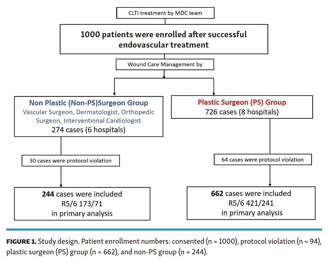
Study design. This study was a multicenter retrospective analysis based on a prospectively maintained database of patients. CLTI patients who successfully underwent percutaneous transluminal angioplasty for isolated infrapopliteal arterial disease at 14 cardiovascular centers from 2006-2012 were enrolled. Their revascularization strategies were left to the discretion of each institution after discussions with their vascular surgeon, interventional radiologist, and cardiologist. A total of 1000 consecutive cases with ischemic tissue loss underwent primary EVT for chronic infrapopliteal arterial ischemia. Forty-six cases with missing follow-up data and 48 cases with missing baseline data of interest were excluded. Finally, 906 cases were included in the primary analysis (Figure 1). This study was a subanalysis based on the database of the J-BEAT (Japanese BElow-the-knee Artery Treatment) registry, which was registered in the University Hospital Medical Information Network Clinical Trial Registry (UMIN-CTR; No. UMIN000004917). The institutional review boards of the participating institutions approved the study. This study was conducted in accordance with the Declaration of Helsinki, and all patients gave written informed consent for both EVT and inclusion in this study prior to the procedure.

EVT protocol. All EVT procedures were performed by EVT specialists, which included cardiovascular interventionists, interventional radiologists, and vascular surgeons. Medications (including pre- and postprocedure aspirin, clopidogrel, ticlopidine, warfarin, and heparin) were administered according to local hospital policy and physician discretion. All EVT procedures were performed under local anesthesia. Angiography was performed via an antegrade approach by ipsilateral common femoral artery access. A guidewire was placed across the lesion and dilated for at least 60 seconds with an optimally sized balloon. Procedures that used a drug-coated balloon, atherectomy device, or stent implantation were excluded from this study because these devices are in non-approval status.
Wound management. Specialists for wound care management were determined by the policy of each hospital. Wound care was provided by plastic surgeons (the PS group; n = 662) at 8 sites, and by vascular surgeons, dermatologists, orthopedic surgeons, and/or interventional cardiologists (the non-PS group; n = 244) at 6 sites. At these participating sites, there are no podiatrists responsible for ischemic wound care. Ischemic ulcers were evaluated and treated during follow-up by either the plastic surgeon(s) or wound care specialists mentioned above.
Wound severity and presence of infection were assessed by the responsible physicians. They also determined indications for antibiotic therapy, wound closure, debridement, dressing, vacuum-assisted closure therapy, and/or hyperbaric oxygen therapy. The timing of repeat revascularization and major or minor amputation was determined when wound healing could not be observed and/or conditions deteriorated. The ulcer status at 1 month and 4 months and the complete ulcer healing time were recorded.
Follow-up protocol. All patients were followed at 1 week and 1 month after revascularization, and then monthly for a total of 4 months, as previous studies had shown complete wound healing to take 3 months on average.5 Follow-up examinations included limb status, ankle-brachial index (ABI), and skin perfusion pressure (SensiLase PAD 3000; Väsamed) measured on the dorsal and plantar surfaces of the foot, when possible.
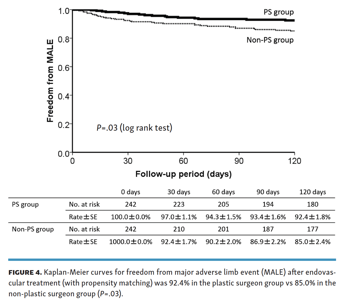
Outcomes. The primary outcome was the comparison of complete wound healing rates between the PS and non-PS groups. Complete healing was defined as the achievement of complete epithelialization of all wounds without any major amputation. To minimize differences in the baseline characteristics of the 2 groups, propensity-score matching was employed and the data were analyzed. The secondary outcomes were a comparison of freedom from major adverse limb event (MALE) rate, defined as a composite of major amputations and surgical conversions between the PS and non-PS groups.
Definitions. Objective ischemic tissue loss has been defined as tissue loss associated with an ankle pressure of <70 mm Hg or a toe pressure of <50 mm Hg.1 When these criteria could not be met, the skin perfusion pressure was measured, and values of <40 mmHg were defined as indicating ischemic tissue loss.
Wounds were classified according to the Rutherford classification.12 Rutherford category 5 denotes minor tissue loss, ie, a non-healing ulcer or focal gangrene with diffuse pedal ischemia. Rutherford category 6 denotes major tissue loss extending above the transmetatarsal level of the functional foot, with the limb no longer salvageable, or located at the heel.
Isolated infrapopliteal artery disease was defined as critical limb ischemia secondary to below-the-knee lesions without any significant popliteal, femoral, iliac, or aortic artery lesions. Patients who died before complete wound healing were counted as delayed wound healing, with the date of death as the cut-off date. In patients who underwent major amputation, the healing time was considered to be infinite.
Statistical analysis. Data are presented as mean ± standard deviation for continuous variables and number (percentage) for discrete variables, unless otherwise stated. A P-value of <.05 was considered statistically significant, and 95% confidence intervals (CIs) were calculated when required. Incidence rates were estimated by the Kaplan-Meier method, and intergroup differences were assessed by the log-rank test. To minimize intergroup differences in baseline characteristics, comparisons between the PS and non-PS groups were performed after propensity-score matching. The propensity score was developed using the logistic regression model in which the following were included as explanatory variables: age, gender, ambulatory status, body mass index, hypertension, hyperlipidemia, diabetes mellitus, chronic renal failure, regular dialysis, smoking status, coronary artery disease, cerebrovascular disease, aspirin use, thienopyridine use, cilostazol use, serum albumin levels, hemoglobin levels, left ventricular ejection fraction, Rutherford classification, detailed wound localization (toe, heel, ankle, sole, and instep), TransAtlantic Inter-Society Consensus (TASC) classification, direct revascularization (defined as angiosome-oriented direct revascularization by which the angiosome-based arteries were recanalized), and below-the-ankle run-off. According to recommendations by Austin et al,17 we matched on the logit of the propensity score with calipers of width equal to 0.2 of the standard deviation of the logit of the propensity score. Intergroup differences after matching were tested as paired analyses. Propensity-score matching was performed with R, version 3.1.0 software (R Foundation for Statistical Computing) and other statistical analyses with SPSS Statistics, version 22 (IBM).
Results
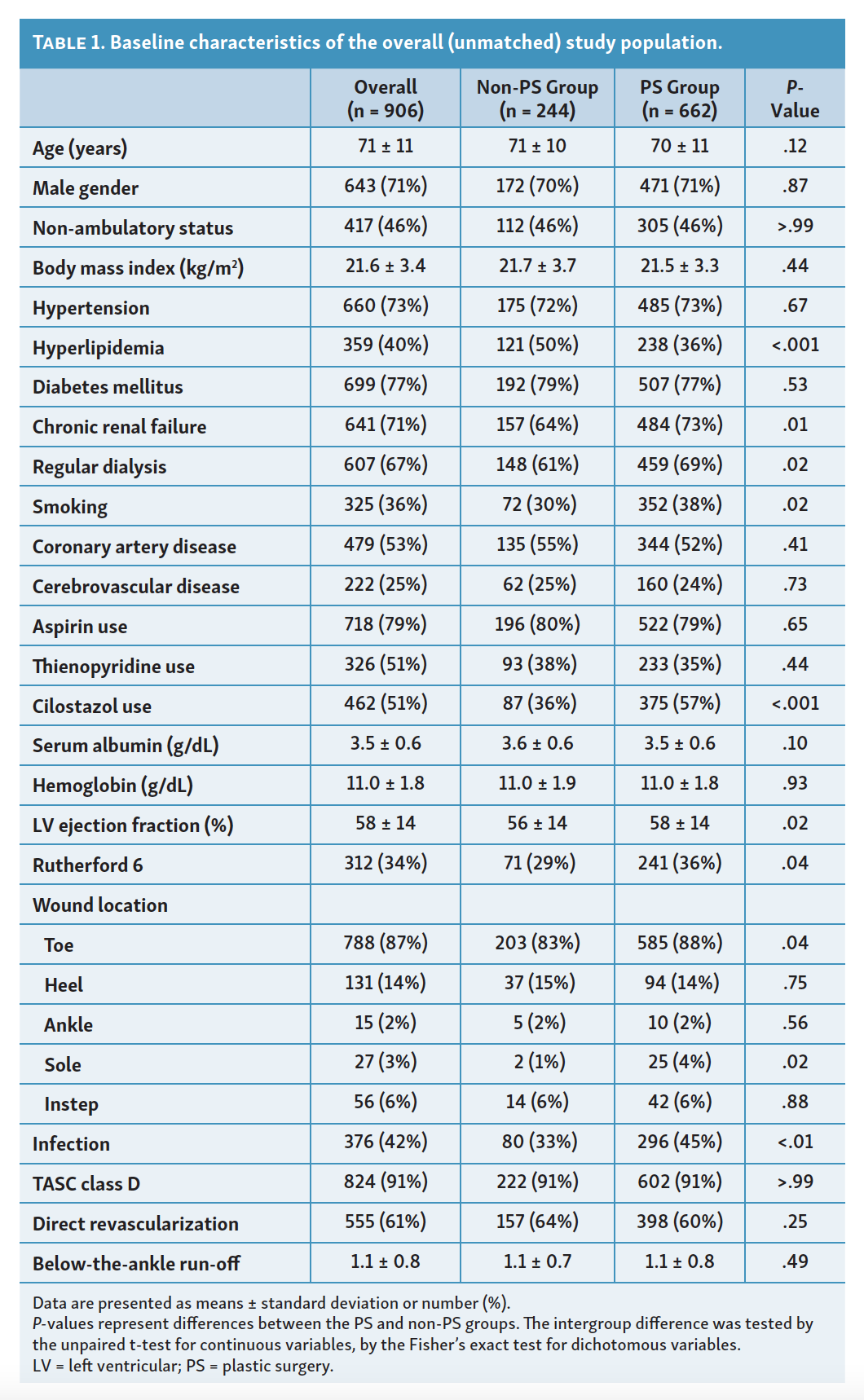
The main clinical characteristics of the included studies are outlined in Table 1. Common clinical characteristics were hypertension (73%), hyperlipidemia (40%), diabetes mellitus (77%), and current smoking (36%). End-stage renal dysfunction on hemodialysis was observed in 607 cases (67%) and Rutherford category 6 disease was noted in 312 cases (34%). A total of 376 cases (42%) were complicated with infection.
The PS group had a higher prevalence of renal dysfunction, history of smoking, Rutherford category 6 symptoms, infection, and more frequent use of cilostazol, whereas the non-PS group was more likely to have hyperlipidemia and lower left ventricular ejection fractions.
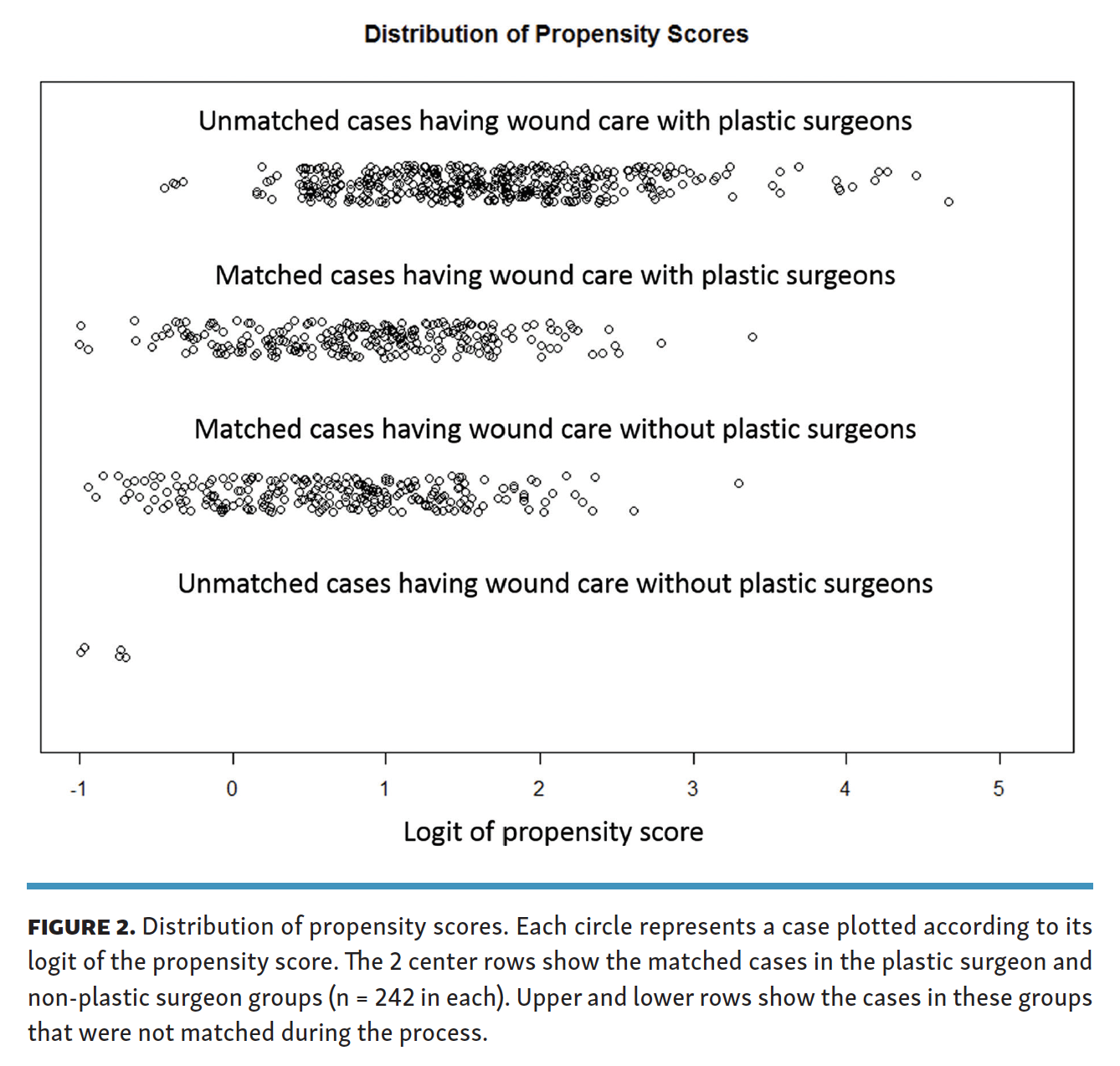
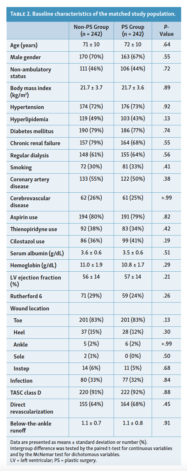
Propensity-score matching extracted 242 pairs from the 2 groups (Figure 2), no significant intergroup difference was found in baseline characteristics (Table 2). As shown in Figure 3, the PS group had a significantly higher wound-healing rate compared with the non-PS group (P=.02), ie, the rate at 4 months was 52.0% (95% CI, 45.3-58.7) vs 40.0% (95% CI, 33.5-46.5%). Moreover, the PS group showed significantly higher freedom from MALE (92.4% vs 85.0%; P=.03) (Figure 4).


Discussion
This study examined the role of plastic surgeons on wound healing after EVT in a cohort of CLTI patients with isolated infrapopliteal lesions. After propensity matching of patient and lesion baseline characteristics, the analysis showed better wound healing in the PS group than in the non-PS group.
In the last decade, EVT procedures for CLTI have been established, and the number of major amputations has been decreasing.5,6 Furthermore, there is a dissociation between amputation-free survival and complete wound healing, and a few patients could not achieve complete wound healing after successful revascularization.2,5 Revascularization is a prerequisite for the effective treatment of CLTI and, combined with careful wound management, is a must for complete ulcer healing.
Recently, physicians and medical staff of different backgrounds have participated in the formation of MDC teams, and several articles have advocated this method to improve CLTI outcomes.11,14,15 Although previous reports have shown the MDC team’s effectiveness, the individual specialists’ roles were not discussed. Because of the rapid development of endovascular revascularization, many teams have experts in revascularization, but not in wound care specific to CLTI. The situation depends on each country. In the European Union, Ukraine, and Australia, the podiatrist takes the role. In other areas, the process is not popular. In Japan, the effectiveness of EVT procedures and wound care by plastic surgeons adept in ischemic wounds have both been demonstrated.16,17 Most patients who need wound care are referred to a plastic surgeon. However, there is a shortage of plastic surgeons specializing in ischemic wound care management despite the great demand for such specialists. In facilities where a plastic surgeon is not available, a team approach consisting of a vascular surgeon, dermatologist, orthopedist, and interventional cardiologist is taken.
No comparative studies of wound-healing rates between plastic surgeons and other medical specialists have been conducted.
There are three reasons for the lack of such data. First, EVT procedures have yet to be standardized and, without the normalization and standardization of EVT treatment strategies, accurate conclusions cannot be reached. However, recent advances have been made in standardization and the 14 facilities that participated in this study were highly skilled and experienced centers with thorough and complete patient evaluations and high success rates. Thus, it can be said that a relatively consistent and standard EVT treatment regimen and appropriate analyses were performed for this study.
Second, the standardization of wound care management has not been established. Plastic surgeons could provide the best treatment options, particularly in complex ischemic wounds. They are familiarized with infection control, wound closure, debridement, dressing, VAC therapy, hyperbaric oxygen therapy, and low-density lipoprotein apheresis therapy.18-22 They could also accurately suggest the timing of minor surgery, amputation, and/or the need for additional revascularization.23,24 The healing rate is significantly lower in patients with PAD complicated by infection, and 42% were complicated by infection in the present study. Even when revascularization was performed, healing was difficult in the presence of infection. Therefore, local control of gangrene and ulcers was indispensable and close cooperation with plastic surgeons was vital.25 However, due to the shortage of plastic surgeons, such wound care is presently being performed by various medical specialists.
Finally, in the current study, the PS group included more severe wounds than in the non-PS group, ie, Rutherford category 6 ischemia and infected limbs. However, in matched analysis, the time course of complete wound healing was faster in the PS group than in the non-PS group. The 4-month wound-healing rate in the PS group was 52% and freedom from MALE was extremely high at 92.4%. This compares favorably with the current state of CLTI treatment, which has a 20% mortality rate at 6 months and a 25%-30% major amputation rate at 1 year. These facts suggest that the specialized care provided by plastic surgeons resulted in more favorable wound-healing outcomes.
To the best of our knowledge, this is the first report comparing ischemic wound care between plastic surgeons adept with ischemic wounds and other medical specialists after EVT. Revascularization alone is insufficient for complete wound healing, and specialized wound care by plastic surgeons or podiatrists was shown to be indispensable for complete healing. MDC teams who are involved after CLTI revascularization are recommended to work together with plastic surgeons or podiatrists for improved rates of complete wound healing.
Study limitations. There were several limitations to this study that may have affected the clinical outcomes. First, this was a retrospective analysis; despite the use of data from multiple large-scale prospective databases and propensity-matched scores, a chance of bias remained in many of the data assignments. Second, data were lacking from the latest classification schemes, the Wound, Ischemia, and Foot Infection (WIfI)26 and Global Anatomic Staging System (GLASS).27 Third, the data presented in this study were mainly from revascularization physicians, and there was a lack of detailed data on wound-management methods. In addition to appropriate revascularization, the types of wound management are also important topics for future investigations.
Conclusion
Our cohort data showed that the rate of complete wound healing after successful EVT in CLTI patients treated by plastic surgeons was superior to the rate for those treated by other medical specialists. These data clearly suggest that revascularization alone is insufficient for complete healing in ischemic wounds. Specialists who focus on ischemic wounds are better equipped to perform wound care treatment than those who do not fully comprehend the complexities of these patients and utilize the emerging technologies that are available to manage them.
Affiliations and Disclosures
From the 1Department of Cardiology, Kishiwada Tokushukai Hospital, Osaka, Japan; 2Department of Diabetes Care Medicine, Osaka University Graduate School of Medicine, Osaka, Japan; 3Cardiovascular Center, Kansai Rosai Hospital, Amagasaki, Japan; 4Department of Cardiology, Kokura Memorial Hospital, Kitakyushu, Japan; 5Department of Vascular Surgery, Matsuyama Red Cross Hospital, Matsuyama, Japan; and the 6Division of Cardiology, Tokyo Rosai Hospital, Tokyo, Japan.
Disclosure: The authors have completed and returned the ICMJE Form for Disclosure of Potential Conflicts of Interest. The authors report no conflicts of interest regarding the content herein.
Manuscript accepted November 29, 2021.
Address for correspondence: Masahiko Fujihara, MD, Kishiwada Tokushukai Hospital Department of Cardiology, 4-27-1 Kamori-cho, Kishiwada City Osaka 596-8522, Japan. Email: masahiko-fujihara@themis.ocn.ne.jp